Recent Advances of Ocular Drug Delivery Systems: Prominence of Ocular Implants for Chronic Eye Diseases
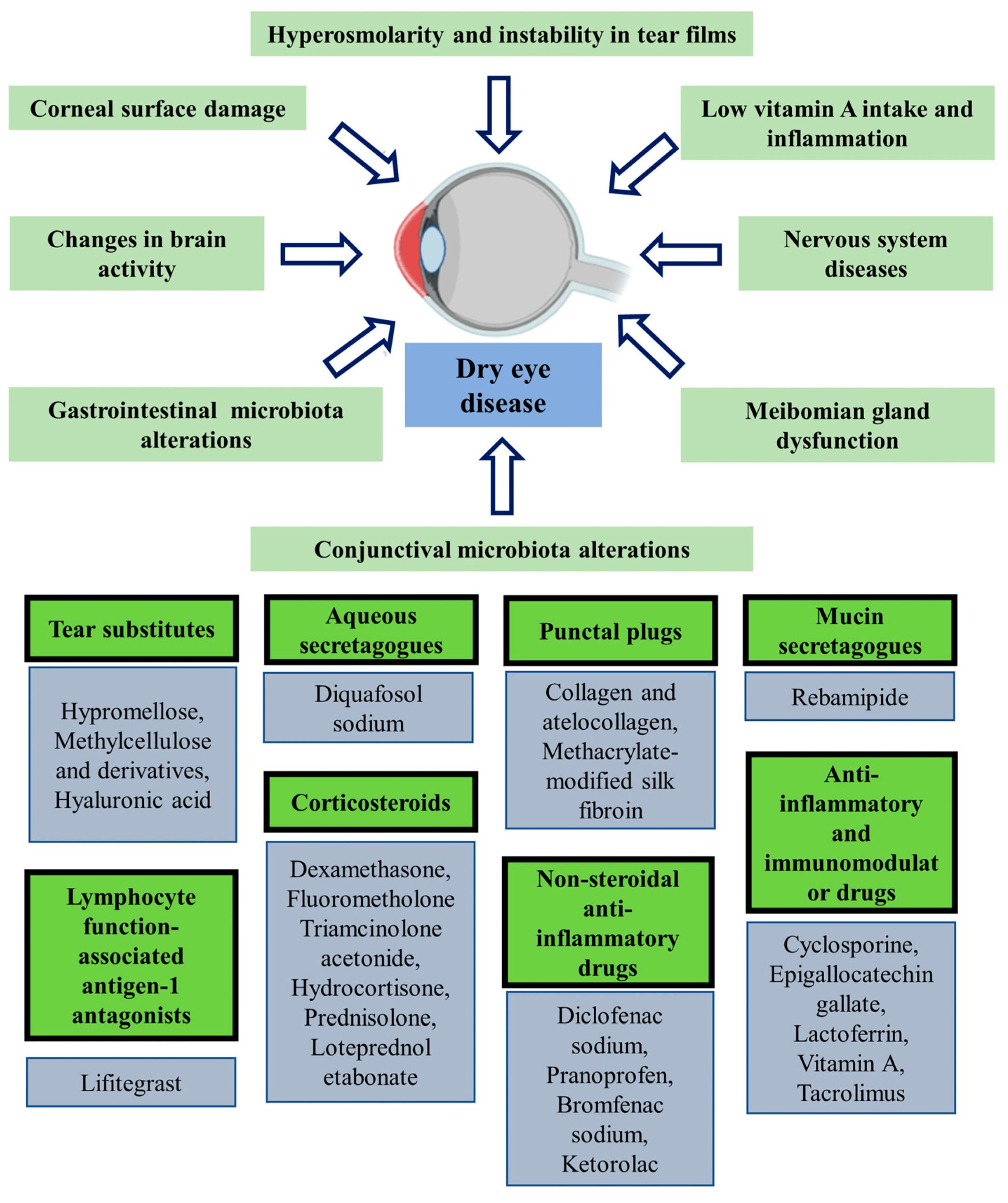
Chronic ocular diseases can seriously impact the eyes and could potentially result in blindness or serious vision loss. According to the most recent data from the WHO, there are more than 2 billion visually impaired people in the world. Therefore, it is pivotal to develop more sophisticated, long-acting drug delivery systems/devices to treat chronic eye conditions. This review covers several drug delivery nanocarriers that can control chronic eye disorders non-invasively. However, most of the developed nanocarriers are still in preclinical or clinical stages. Long-acting drug delivery systems, such as inserts and implants, constitute the majority of the clinically used methods for the treatment of chronic eye diseases due to their steady state release, persistent therapeutic activity, and ability to bypass most ocular barriers. However, implants are considered invasive drug delivery technologies, especially those that are nonbiodegradable. Furthermore, in vitro characterization approaches, although useful, are limited in mimicking or truly representing the in vivo environment. This review focuses on long-acting drug delivery systems (LADDS), particularly implantable drug delivery systems (IDDS), their formulation, methods of characterization, and clinical application for the treatment of eye diseases.

1. Introduction

Over the last three decades, the prevalence of sight-related diseases has received increased attention; this is mainly due to the increasing life expectancy of the global population. There were around 188 million people who had minor vision impairment, 216 million people who had moderate-to-severe sight impairment, and approximately 40 million people who were legally blind [1]. These numbers are only expected to increase over time. The eye has a complicated vital structure with several anatomical and physiological constraints. The anterior part of the eye, which is implicated in refraction and vision, is made up of several ocular tissues, including the cornea, conjunctiva, aqueous humor, iris, ciliary body, and the lens, whereas the back segment of the eye is mostly made up of the vitreous humor, choroid, retina, and posterior sclera. The posterior segment recognizes and transmits light signals though the optic nerve so that the eye can view the outside world. Many chronic eye diseases can affect these specialized ocular tissues.
Common conditions that affect the front of the eye include glaucoma, anterior uveitis, cataracts, and dry eye diseases [2,3], while the conditions that most often affect the back of the eye include AMD, diabetic retinopathy (DR), CMV, vitreoretinopathy, and posterior uveitis [3]. Topical eye drops supply drugs to most of anterior segment tissues, whilst eye injections (most notably, intravitreal) are the standard drug administration option for posterior segment diseases. Poor bioavailability (less than 5%) represents a major issue with topically administered ocular medications, while invasiveness (typically repeated monthly intravitreal injections) and non-compliance issues are the main hurdles to treating the diseases of the posterior segment.
The barriers to treating diseases of the anterior segment include a tight corneal–epithelial junction, reflex blinking and tearing, ocular tissue/s metabolism, tear turnover, nasolacrimal drainage, efflux transporter pumps, and the blood–aqueous barrier [4]. These anatomical and physiological constraints have been discussed in detail elsewhere [3,5]. The main barrier to medication absorption following topical application is the corneal epithelium. Tight intercellular connections surround cells that are on the surface serve as barriers to prevent drug molecules from entering the cells through the paracellular route [6]. The typical drop size of topically instilled eye drops, which is delivered to the eye, is in the volume range of 25–56 µL. Although the human eye can temporarily accommodate up to 30 µL, any excess amount is quickly wasted due to reflex blinking, greatly reducing the amount of medication that is ultimately accessible for a therapeutic effect [7,8]. P-glycoprotein and multidrug resistant proteins primarily cause drug efflux. P-glycoprotein, which is located in the blood–aqueous and blood–retinal barriers [9,10], eliminates amphipathic substances, while multidrug resistant proteins, which are found in the ciliary body and blood–aqueous barrier [11], are known to export organic anions. Endothelial cells from the blood vessels in the iris and cilia form the blood–aqueous barrier together with the non-pigmented ciliary epithelium. This prevents the bulk of medications from reaching deeper ocular tissues and controls the diffusion of soluble molecules between the front and back of the eye by building tight connections at the cellular level [12,13].
For drugs targeted at the back of the eye, the retinal pigmented epithelium, ciliary body, and ocular metabolic enzymes reduce how much of the drug remains [13,14]. The posterior segment barriers include the inner limiting membrane, vitreous diffusion, tight retinal–pigmented epithelium junctions, and the blood–retinal barrier [15]. The inner limiting membrane is a substantial physical barrier that inhibits drugs from being delivered to the posterior portion of the eye [16]. The vitreous body represents a second major barrier for drug delivery for the posterior segment. In the human eye, the vitreous body, a transparent, gel-like substance, accounts for around 80% of the total volume. The vitreous body consists of extraordinarily high water content (>97%) and collagen fibers [17,18]. The collagen fibers make up the network that fabricate the gel structure in the 3D shape and make it flexible and strong against mechanical pressures. The vitreous body can act as a barrier either physiologically or anatomically. The physiological barrier action is represented by the slowing down of drug diffusion and the anatomical barrier is represented by the 3D gel-like structure [19]. The retina and retinal pigment epithelium’s limiting structure prevents the free flow or diffusion of therapeutic drugs, which is what gives the retina its tight junctions [20]. Another significant challenge to drug delivery to the posterior portion is the blood–retinal barrier. The outer and inner blood–retinal barriers make up the blood–retinal barrier. The inner blood–retinal barrier is made up of retinal capillary endothelial cells, whereas the outside blood–retinal barrier is composed of tightly connected retinal pigment epithelial cells. Similar to the blood–brain barrier, the absence of wide gaps in the retinal pigment epithelium and retinal endothelial cells prevents passive drug transport. Only very small molecules from the choroid, including carbon dioxide, oxygen, and lipophilic compounds, can diffuse to the inner retinal tissues [21].
In this review, the most common ocular chronic disorders will be discussed. These conditions necessitate longer treatment intervals with drugs, and the most effective drug delivery systems should ideally improve the activity, stability, and distribution of drug molecules to target the ocular tissues. The utilization of long-acting drug delivery systems (LADDS), particularly implantable drug delivery systems (IDDS), and their formulation and methods of characterization, assessment, and their clinical application are covered.


Download the full review as PDF here: Recent Advances of Ocular Drug Delivery Systems: Prominence of Ocular Implants for Chronic Eye Diseases

or read it here 

Mostafa, M.; Al Fatease, A.; Alany, R.G.; Abdelkader, H. Recent Advances of Ocular Drug Delivery Systems: Prominence of Ocular Implants for Chronic Eye Diseases. Pharmaceutics 2023, 15, 1746.
https://doi.org/10.3390/pharmaceutics15061746

You might also like